X-rays have revolutionized the field of medical imaging, providing valuable insights into the human body’s inner workings. Discovered in 1895 by German physicist Wilhelm Conrad Roentgen, X-rays are a form of electromagnetic radiation that possess high energy and short wavelengths. They have since become an essential tool in medicine, offering non-invasive and detailed images of bones, tissues, and organs. In this comprehensive discussion, we will explore the principles behind X-ray technology, its historical significance, its applications in various fields, and the safety considerations associated with its use.
X-rays, X-rays, X-rays—these electromagnetic waves have become synonymous with diagnostic imaging. They belong to the electromagnetic spectrum, which encompasses a range of waves with varying wavelengths and energies. X-rays, specifically, lie between ultraviolet radiation and gamma rays in this spectrum. Their high energy and short wavelength enable them to penetrate solid objects, making them highly useful in medical imaging.
Wilhelm Conrad Roentgen’s discovery of X-rays in 1895 was a momentous event in scientific history. While experimenting with cathode rays, Roentgen noticed that a fluorescent screen in his laboratory began to glow even when it was positioned far from the cathode tube. He deduced that a new type of radiation was responsible for this phenomenon and named it X-ray, with the “X” symbolizing the unknown nature of this radiation. Roentgen’s groundbreaking work earned him the first-ever Nobel Prize in Physics in 1901, cementing the significance of X-rays in the scientific community.
X-ray technology quickly found its way into the medical field, where its applications proved to be invaluable. One of the earliest medical uses of X-rays was in the imaging of skeletal structures. By passing X-ray beams through the body and capturing the resulting images on photographic film, doctors gained the ability to visualize bones and diagnose fractures, dislocations, and other skeletal abnormalities. This development revolutionized orthopedics, as it enabled doctors to accurately assess bone injuries without invasive procedures.
In addition to skeletal imaging, X-rays have proven to be instrumental in the detection and diagnosis of various diseases and conditions. X-ray images can reveal the presence of tumors, infections, and other abnormalities within organs and soft tissues. This has been particularly useful in fields such as radiology, oncology, cardiology, and gastroenterology, where X-rays are routinely employed to aid in diagnoses and treatment planning.
The process of obtaining X-ray images, known as radiography, involves several key components. X-ray machines consist of an X-ray tube that generates the X-ray beam and a detector or film that captures the transmitted radiation. When a patient undergoes an X-ray examination, they are positioned between the X-ray tube and the detector. The X-ray machine is then activated, emitting a controlled amount of radiation that passes through the body. The X-rays that are not absorbed by the body’s tissues reach the detector, which converts them into a visible image. This image can be digitally displayed on a computer screen or recorded on a photographic film.
Safety considerations are of utmost importance when it comes to X-ray imaging. While X-rays are incredibly useful, they can also pose risks if not properly managed. Prolonged or excessive exposure to X-rays can damage living tissues and potentially lead to adverse health effects, such as radiation burns, DNA damage, and an increased risk of cancer. As a result, strict safety protocols and regulations are in place to ensure that patients and medical personnel are protected from unnecessary radiation exposure.
To minimize radiation exposure, various measures are implemented during X-ray procedures. Lead aprons and shields are commonly used to protect areas of the body that are not being imaged from unnecessary radiation. Collimation, which involves restricting the size and shape of the X-ray beam, helps to focus the radiation on the specific area of interest and reduces scatter radiation. Additionally, modern X-ray machines are designed to emit the lowest possible amount of radiation required to obtain diagnostically useful images.
Advancements in technology have further expanded the applications of X-rays beyond traditional radiography. Computed tomography (CT) scans, for instance, utilize X-ray beams and sophisticated computer algorithms to create detailed cross-sectional images of the body. CT scans provide three-dimensional views and are particularly valuable in diagnosing conditions related to the brain, chest, abdomen, and pelvis. They are also useful for guiding interventional procedures, such as biopsies and drainages.
Another innovative application of X-rays is in the field of mammography, which focuses on breast imaging. Mammograms employ low-dose X-rays to detect breast cancer at an early stage, allowing for prompt treatment and increased chances of survival. This screening technique has proven to be highly effective in reducing breast cancer mortality rates, emphasizing the life-saving potential of X-ray technology.
Outside of medicine, X-rays find utility in various industries and fields of research. In materials science, X-ray diffraction is a powerful technique used to determine the atomic and molecular structures of crystals. This method has contributed significantly to the understanding of the arrangement and properties of materials, aiding in the development of new alloys, pharmaceuticals, and electronic devices.
Archaeology and paleontology also benefit from X-ray analysis. By examining the internal structures of fossils and artifacts, researchers can gain insights into their composition, preservation, and evolutionary significance. X-ray imaging techniques have uncovered hidden details and shed light on ancient civilizations, extinct species, and archaeological mysteries.
Beyond their applications in medical imaging, X-rays have found utility in numerous other fields, ranging from security and industry to astronomy and art conservation. Let’s delve further into these diverse applications and explore the ongoing developments in X-ray technology.
Industrial applications of X-rays are also widespread, particularly in non-destructive testing (NDT). X-ray inspection techniques are utilized to assess the integrity and quality of manufactured components, such as welds, pipelines, and aircraft parts. By subjecting these objects to X-ray scrutiny, any structural defects, cracks, or inconsistencies can be detected, ensuring the reliability and safety of industrial equipment and infrastructure.
Over the years, advancements in X-ray technology have driven the development of more sophisticated imaging modalities. Digital radiography (DR) has replaced traditional film-based X-ray imaging, offering immediate image acquisition and improved image quality. DR systems use digital detectors to capture X-ray images, which can be enhanced, manipulated, and transmitted electronically. This digital approach enhances workflow efficiency, reduces radiation exposure, and enables seamless integration with picture archiving and communication systems (PACS).
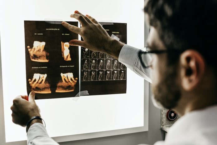
Another significant advancement is the emergence of cone-beam computed tomography (CBCT). While traditional CT scans utilize a fan-shaped X-ray beam, CBCT employs a cone-shaped X-ray beam, resulting in volumetric image acquisition. CBCT is particularly useful in dentistry and maxillofacial imaging, allowing dentists and oral surgeons to visualize complex anatomical structures in three dimensions, aiding in treatment planning for dental implants, orthodontics, and oral surgeries.
Furthermore, X-ray fluorescence (XRF) spectroscopy has emerged as a powerful analytical technique for elemental analysis. XRF involves bombarding a sample with X-rays, which cause the atoms to emit characteristic fluorescent X-rays. By measuring the energy and intensity of these emitted X-rays, scientists can determine the elemental composition of the sample. XRF finds applications in fields such as geology, archaeology, environmental science, and forensics, facilitating the identification and quantification of elements present in various materials.
In recent years, the field of X-ray imaging has witnessed notable advancements in image resolution, speed, and radiation dose reduction. High-resolution X-ray imaging techniques, such as phase-contrast imaging and coherent diffractive imaging, have enabled the visualization of fine structures and subtle density variations in biological tissues and materials. These techniques rely on the interactions of X-rays with matter beyond conventional absorption, providing enhanced contrast and improved image quality.
Furthermore, efforts to minimize radiation exposure during X-ray procedures have led to the development of low-dose imaging techniques. Iterative reconstruction algorithms and advanced image processing techniques have been implemented to reduce the radiation dose required while maintaining diagnostically useful image quality. Such innovations prioritize patient safety by balancing the need for accurate diagnosis with minimizing potential risks associated with radiation exposure.
Looking ahead, the future of X-ray technology holds great promise. Ongoing research aims to further refine imaging modalities and develop new applications. For instance, X-ray phase-contrast tomography, which exploits the phase shifts of X-rays passing through different tissues, shows potential for improved soft tissue imaging, enabling better differentiation of structures with similar X-ray absorption properties.
Moreover, the combination of X-ray imaging with other imaging modalities, such as magnetic resonance imaging (MRI) or positron emission tomography (PET), has the potential to provide comprehensive and complementary information about the body’s structure and function. These multimodal imaging approaches could enhance diagnostic accuracy, aid in personalized medicine, and contribute to our understanding of complex diseases.
In conclusion, X-ray technology has had a profound impact across various fields, transcending its initial application in medical imaging. From aiding in the diagnosis and treatment of diseases to enhancing industrial inspections, ensuring security, and uncovering the secrets of the cosmos, X-rays continue to be an indispensable tool for scientific inquiry and practical applications. As technology advances and our understanding of X-ray interactions with matter deepens, the potential for further breakthroughs and innovations in X-ray imaging remains vast, holding the promise of continued advancements in healthcare, industry, and scientific exploration.


